Slip Disc: How Ayurveda & Panchkarma are beneficial

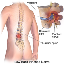
Spinal disc herniation, also known as a slipped disc, is a medical condition affecting the spine in which a tear in the outer, fibrous ring of an intervertebral disc allows the soft, central portion to bulge out beyond the damaged outer rings.

Disc herniation is usually due to age-related degeneration of the outer ring, known as the anulus fibrosus, although trauma, lifting injuries, or straining have been implicated as well. Tears are almost always postero-lateral (on the back of the sides) owing to the presence of the posterior longitudinal ligament in the spinal canal. This tear in the disc ring may result in the release of chemicals causing inflammation, which may directly cause severe pain even in the absence of nerve root compression.

Disc herniations are normally a further development of a previously existing disc protrusion, a condition in which the outermost layers of the anulus fibrosus are still intact, but can bulge when the disc is under pressure. In contrast to a herniation, none of the central portion escapes beyond the outer layers. Most minor herniations heal within several weeks. Anti-inflammatory treatments for pain associated with disc herniation, protrusion, bulge, or disc tear are generally effective. Severe herniations may not heal of their own accord and may require surgery. The condition may be referred to as a slipped disc, but this term is not accurate as the spinal discs are firmly attached between the vertebrae and cannot “slip” out of place.

Symptoms

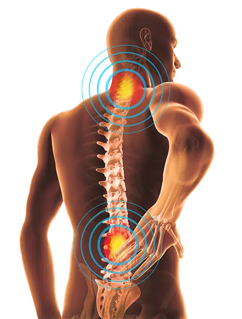
Symptoms of a herniated disc can vary depending on the location of the herniation and the types of soft tissue that become involved. They can range from little or no pain if the disc is the only tissue injured, to severe and unrelenting neck pain or low back pain that will radiate into the regions served by affected nerve roots that are irritated or impinged by the herniated material. Often, herniated discs are not diagnosed immediately, as the patients come with undefined pains in the thighs, knees, or feet.

Other symptoms may include sensory changes such as numbness, tingling, paresthesia, and motor changes such as muscular weakness, paralysis, and affection of reflexes. If the herniated disc is in the lumbar region, the patient may also experience sciatica due to irritation of one of the nerve roots of the sciatic nerve. Unlike a pulsating pain or pain that comes and goes, which can be caused by muscle spasm, pain from a herniated disc is usually continuous or at least is continuous in a specific position of the body.

It is possible to have a herniated disc without any pain or noticeable symptoms, depending on its location. If the extruded nucleus pulposus material doesn’t press on soft tissues or nerves, it may not cause any symptoms. A small-sample study examining the cervical spine in symptom-free volunteers has found focal disc protrusions in 50% of participants, which suggests that a considerable part of the population can have focal herniated discs in their cervical region that do not cause noticeable symptoms.

A prolapsed disc in the lumbar spine can cause radiating nerve pain. This type of pain is usually felt in the lower extremities or groin area. Radiating nerve pain caused by a prolapsed disc can also cause bowel and bladder incontinence.

Typically, symptoms are experienced only on one side of the body. If the prolapse is very large and presses on the nerves within the spinal column or the cauda equina, both sides of the body may be affected, often with serious consequences. Compression of the cauda equina can cause permanent nerve damage or paralysis. The nerve damage can result in loss of bowel and bladder control as well as sexual dysfunction. This disorder is called cauda equina syndrome. Other complications include chronic pain.

Causes

Most authors favour degeneration of the intervertebral disc as the major cause of spinal disc herniation and cite trauma as a low cause. Disc degeneration occurs both with degenerative disc disease and aging. With degeneration, the contents of the disc, the nucleus pulposus and anulus fibrosus, are exposed to altered loads. Specifically, the nucleus becomes fibrous and stiff and less able to bear load. The load is transferred to the anulus, which, if it fails to bear the increased load, can lead to the development of fissures. If the fissures reach the periphery of the anulus, the nuclear material can pass through as a disc herniation.

Disc herniations can result from general wear and tear, such as constant sitting or squatting, driving, or a sedentary lifestyle. However, herniations can also result from the lifting of heavy loads.[citation needed] Professional athletes, especially those playing contact sports such as American football, are prone to disc herniations as well. Within athletic contexts, herniation is often the result of sudden blunt impacts against, or abrupt bending or torsional movements of, the lower back. When the spine is straight, such as in standing or lying down, internal pressure is equalized on all parts of the discs. While sitting or bending to lift, internal pressure on a disc can move from 17 psi (lying down) to over 300 psi (lifting with a rounded back). Herniation of the contents of the disc into the spinal canal often occurs when the anterior side (stomach side) of the disc is compressed while sitting or bending forward, and the contents (nucleus pulposus) get pressed against the tightly stretched and thinned membrane (anulus fibrosus) on the posterior side (back side) of the disc. The combination of membrane thinning from stretching and increased internal pressure (200 to 300 psi) results in the rupture of the confining membrane. The jelly-like contents of the disc then move into the spinal canal, pressing against the spinal nerves, which may produce intense and potentially disabling pain and other symptoms.

Several genes are also associated with intervertebral disc degeneration. Probable candidate genes like type I collagen (sp1 site), type IX collagen, vitamin D receptor, aggrecan, asporin, MMP3, interleukin-1, and interleukin-6 polymorphisms have been implicated in disc degeneration. Mutation in genes coding for proteins involved in the regulation of the extracellular matrix, such as MMP2 and THBS2, has been demonstrated to contribute to lumbar disc herniation.


Treatment

Diagnosis is made by a Doctor based on your history, symptoms, a physical examination, and results of tests, including the following:
  • X-ray: Application of radiation to produce a film or picture of a part of the body can show the structure of the vertebrae and the outline of the joints. X-rays of the spine are obtained to search for other potential causes of pain, i.e. tumors, infections, fractures, etc.
  • Computed tomography scan (CT or CAT scan): A diagnostic image created after a computer reads x-rays; can show the shape and size of the spinal canal, its contents, and the structures around it.
  • Magnetic resonance imaging (MRI): A diagnostic test that produces three-dimensional images of body structures using powerful magnets and computer technology; can show the spinal cord, nerve roots, and surrounding areas, as well as enlargement, degeneration, and tumours.
  • Myelogram: An x-ray of the spinal canal following injection of a contrast material into the surrounding cerebrospinal fluid spaces; can show pressure on the spinal cord or nerves due to herniated discs, bone spurs or tumours.
  • Electromyogram and Nerve Conduction Studies (EMG/NCS): These tests measure the electrical impulse along nerve roots, peripheral nerves, and muscle tissue. This will indicate whether there is ongoing nerve damage if the nerves are in a state of healing from a past injury, or whether there is another site of nerve compression.

Ayurveda Management

Ayurveda categorized Slip disc as one of the diseases caused by vitiation Vata (one of the principle dosha in the body, responsible for the movement and functionality of the body). Treatment in Ayurveda is aimed at restoring the equilibrium through correction of the underlying functional in-equilibrium. Ayurvedic treatments for Slip disc concentrate on bringing back the aggravated vata to the state of equilibrium and thereby to the state of health.

At Bhagwati Ayurveda, we are providing very effective and most successful treatment for Slip disc or herniated disc. With our vast experience in treating Spine related conditions, today we are successfully dealing even post-surgical recurrence cases.

The treatment comprises of Detoxification and rejuvenation through Ayurvedic Panchakarma therapy, administration of researched medicines internally.

The therapies like Abhyanga swedam, Nasyam, Pathrapotala swedam, Choornapinda swedam, Pizhichil, Shirodhara, Kadeevasthy, Greevavasthy, Navarakizhi, Vasti etc. are done as per the necessity and condition. These therapies are directed towards relieving the inflammatory changes, releasing the spasms and nerve compressions in the affected area, strengthening the supportive tissues holding the spine, nourishing the joints by improving the circulation. Usually, the treatment period is 4 – 5 weeks according to the severity of the disease.

In four to six weeks, the majority of patients find their symptoms are relieved, without surgery. If patient can come for the treatment in early stages, even total cure without recurrence is also possible.

Prevention

Because there are various causes for back injuries, prevention must be comprehensive. Back injuries are predominant in manual labor so the majority low back pain prevention methods have been applied primarily toward biomechanics. Prevention must come from multiple sources such as education, proper body mechanics, and physical fitness.

Education

Education should emphasize not lifting beyond one’s capabilities and giving the body a rest after strenuous effort. Over time, poor posture can cause the IVD to tear or become damaged. Striving to maintain proper posture and body alignment will aid in preventing disc degradation.

Exercise

Exercises that enhance back strength may also be used to prevent back injuries. Back exercises include the prone press-ups, upper back extension, transverse abdominus bracing, and floor bridges. If pain is present in the back, it can mean that the stabilization muscles of the back are weak and a person needs to train the trunk musculature. Other preventative measures are to lose weight and to not work oneself past fatigue. Signs of fatigue include shaking, poor coordination, muscle burning, and loss of the transverse abdominal brace. Heavy lifting should be done with the legs performing the work, and not the back.

Swimming is a common tool used in strength training. The usage of lumbarsacral support belts may restrict movement at the spine and support the back during lifting.